


Dr. Hermann Aberle
University of Münster (Germany), Institute for Neurobiology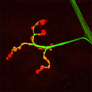
A single nerve branch exits a major motor nerve to innervate a muscle fiber in the lateral body wall region of a Drosophila larva. Immunohistochemical staining using antibodies against the cytoskeletal protein Futsch (green, Alexa Fluor 488) and the Drosophila vesicular glutamate transporter DVGLUT (red, Cy3).
Research Focus & Application:
The research focus of my lab is the establishment and remodeling of synaptic connections between motor neurons and muscle fibers.
Microscopy and Imaging Methods:
We use a ZEISS LSM 510 Meta because of its ability to image multiple colors at high resolution.