


Dr. Jonathan Cohen
National Institute of Child Health and Human Development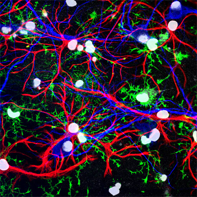
Dissociated hippocampal cultures derived from embryonic day 18.5 rats were fixed and immuno- stained after 12 days in vitro. Neuronal cells were stained with anti-MAP2 (shown in blue), astrocytic cells were stained with anti-GFAP (shown in red), and oligodendrocytic cells were stained with anti-NG2 (shown in green). Cell nuclei are shown in white from staining with Hoescht 33342. These cells were imaged using two photon microscopy.
Research Focus & Application:
My work in the laboratory of R. Douglas Fields is on how neuron-glial interactions and synaptic activity regulate development in the central nervous system. In particular we are focused on how these processes function during learning and plasticity. In these experiments, the mixed hippocampal culture preparation allows us to study how neurons and glia develop and interact through neuron-glial signaling as a model for in vivo development.
Microscopy and Imaging Methods:
I utilize the LSM 510 NLO two-photon confocal microscope in my work as it allows me to image under a wide range of applications including four-color labeling for multiple cell types present in culture, live cell calcium imaging, and fluorescent protein trafficking of cultured cells and thick tissue sections.