


Dr. Hang Hu
Department of Neurobiology & Anatomy, West Virginia UniversitySensory Neuroscience Research Center
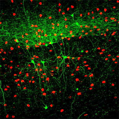
We stained fixed brain sections from somatosensory cortex of GAD67-GFP transgenic mice with antibodies to somatostatin and parvalbumin. We observed that GFP expressing interneurons were colocalized with somatostatin but not with parvalbumin immuno- reactivity. This technique allowed us to classify the somatostatin containing interneurons into subtypes, based on their electro- physiological, morphological and neurochemical properties.
Cells pseudocolored red contain parvalbumin (PV). Cells pseudo- colored green express GFP.
Research Focus & Application:
Dr. Agmon's lab generated transgenic mice in which specific subsets of inhibitory interneurons are induced to express green fluorescent protein (GFP), and are thus easily targetable for electrophysiological recordings in living brain slices. This image was taken by Dr. Hang Hu on the ZEISS LSM 510 confocal microscope system, in the course of research conducted in Dr. Ariel Agmon's laboratory at West Virginia University. Dr. Agmon's research focuses on synaptic circuitry and development in the cerebral cortex with emphasis on thalamocortical connections and feed-forward inhibition.
Dr. Hang Hu (left) and Dr. Agmon at Sensory Neuroscience Research Center,
West Virginia University
Microscopy and Imaging Methods:
We can clearly identify that GFP neurons are labeled with different markers through the multi-fluorescence channels of the LSM 510 confocal microscope.