


Dr. Joshua Zimmerberg
Laboratory of Cellular and Molecular Biophysics at NICHDNational Institutes of Health (NIH)
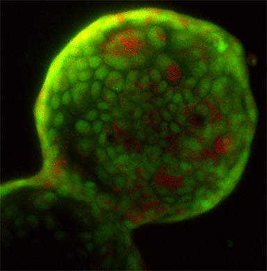
MDCK cells growing on beads are stained with Acridine Orange, which labels live cells in green and acidotic cells in orange.
Research Focus & Application:
The Section on Membrane and Cellular Biophysics, led by Joshua Zimmerberg, studies membrane mechanics, intracellular molecules, membranes, viruses, organelles, and cells to understand viral and parasite infection, exocytosis, and apoptosis. This section has organized an interdisciplinary attack on the mechanisms of membrane remodeling with varied techniques and approaches, using both the physics of continuum bilayers and direct observations of biological fusion, analytical and numerical calculations of membrane energetics, and experiments on phospholipid bilayers, purified proteins, cell expression systems, purified organelles, cell surface complexes, and the actual physiological and pathogenic events of fertilization and viral infection.
Microscopy and Imaging Methods:
Confocal Microscopy on the ZEISS 510 allows us to image live and dead cells and coverage simultaneously with one dye only. Acridine orange stains live cells green and acidotic cells orange. Comparison to the brightfield or DIC image shows how much of the bead is covered with cells.