


Dr. Albert Ayoub
Department of Neurobiology / Yale University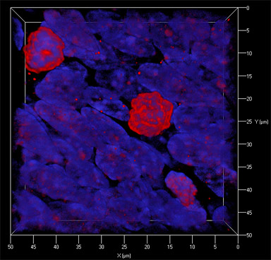
Embryonic SVZ cell (neuronal progenitor) nuclei stained with Ki67 (red) and DAPI (blue).
Research Focus & Application:
The study of neural stem cell subtypes in human and animal brain tissue using multiple Immunohistochemistry.
Microscopy and Imaging Methods:
Multichannel fluorescence and three-dimensional reconstructions of neural stem cells using an Axioplan 2 microscope, Apotome, Axiocam MRm and 40x (0.9 Korr) Plan-Apo. We used 3D Deconvolution and Inside4D to reconstruct and rotate the stack. We use ZEISS because it provides the best optical resolution in its class, advanced 3D rendering capabilities, user friendliness and reliability.