


The in vitro culture of whole tissues and isolated cells was first undertaken in the early 1900s as a technique for investigating the behavior of normal animal cells in an isolated and highly controlled environment. The term tissue culture arose because most of the early cell lines were derived from primary tissue explants, a technique that dominated the field for over 50 years. As established cell lines that can be subcultured and preserved by freezing have slowly emerged, the application of well-defined normal and transformed cells in a host of biomedical investigations has become an important staple in the advanced development of cellular and molecular biology. This fluorescence image gallery explores a number of cell lines labeled with a variety of fluorophores using both traditional staining methods as well as immunofluorescence techniques.
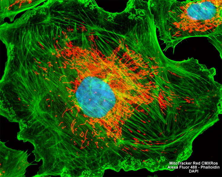
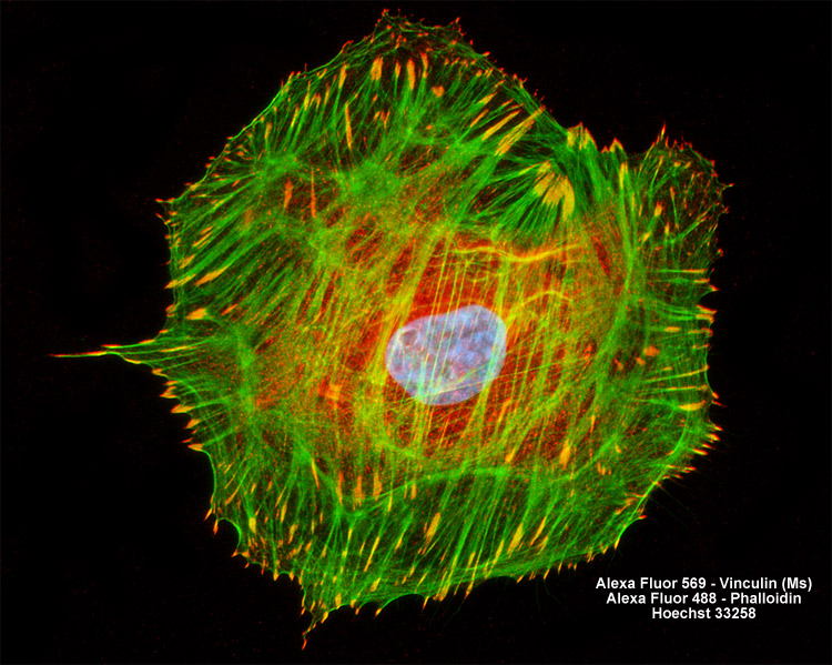
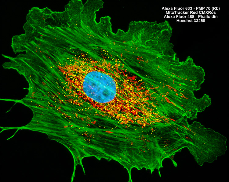
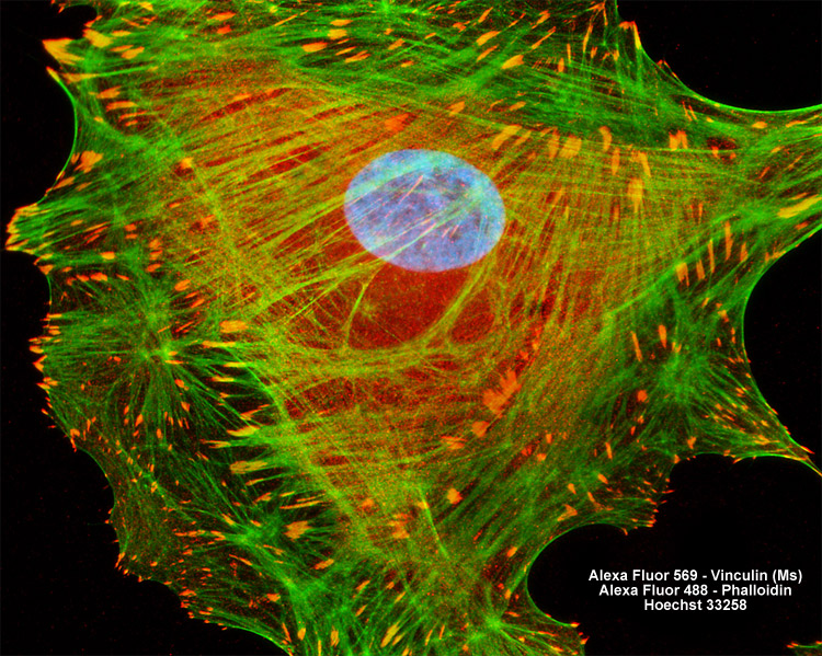
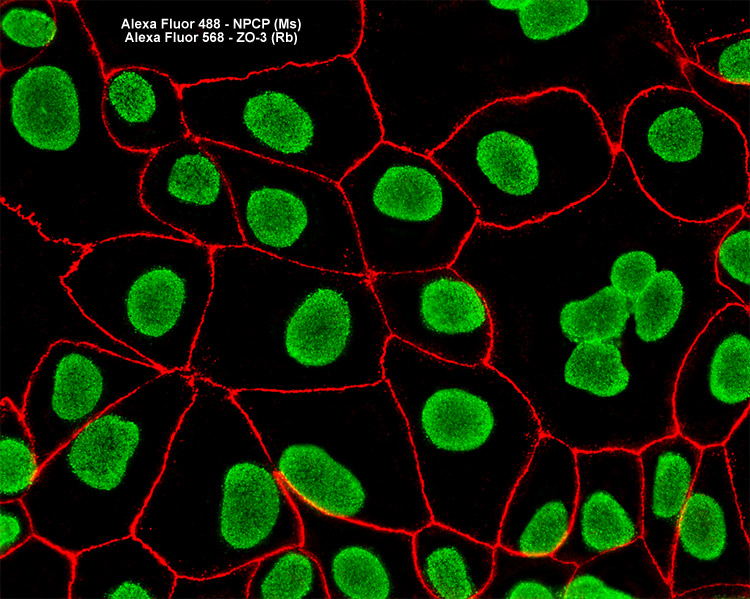
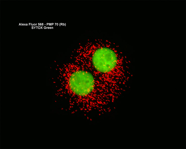
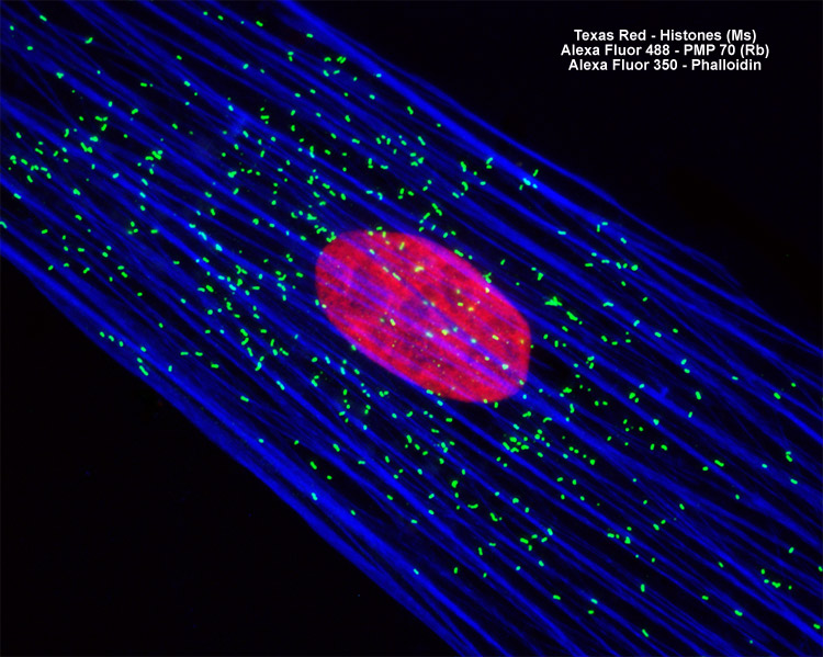

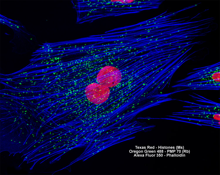
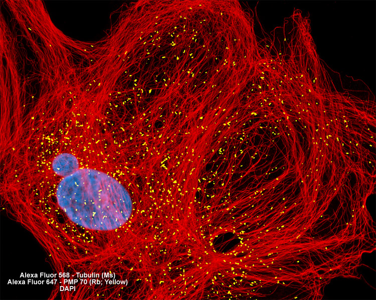
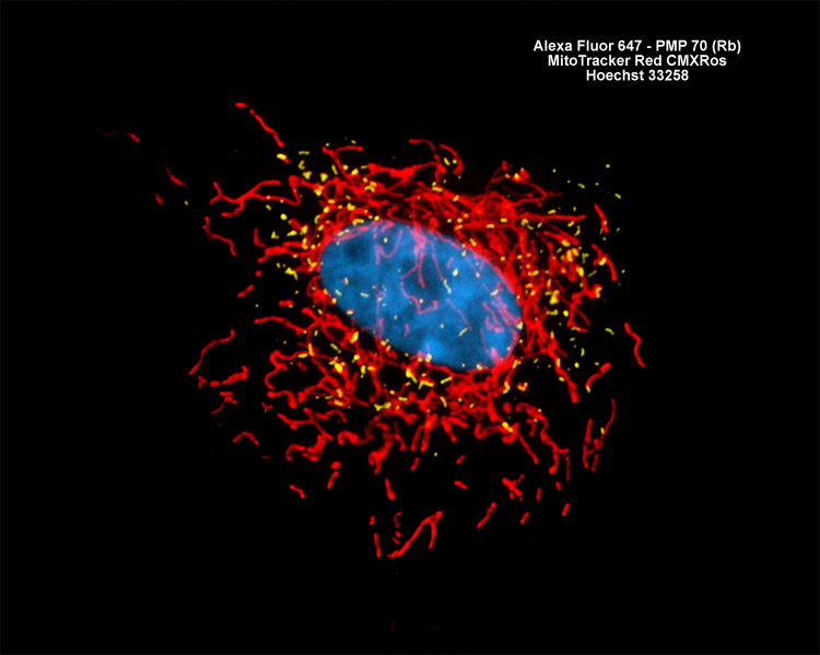

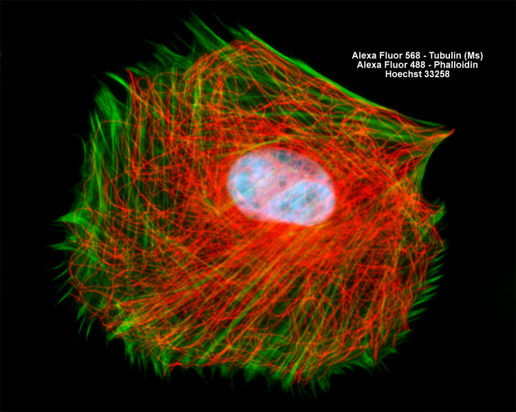
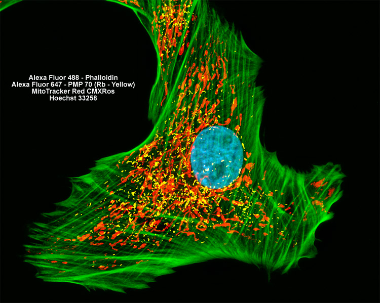
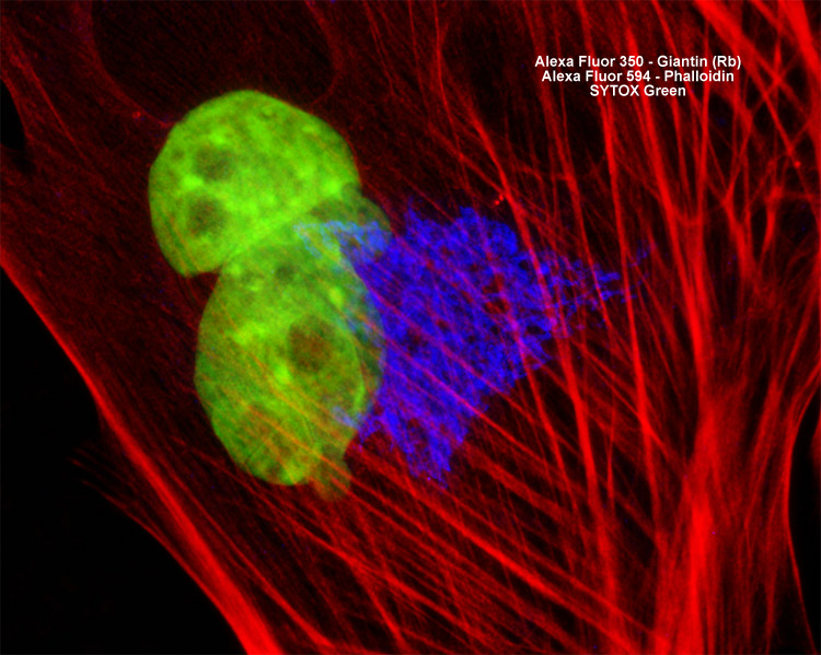
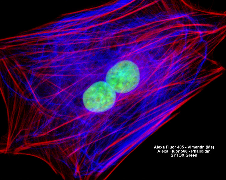
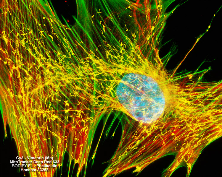
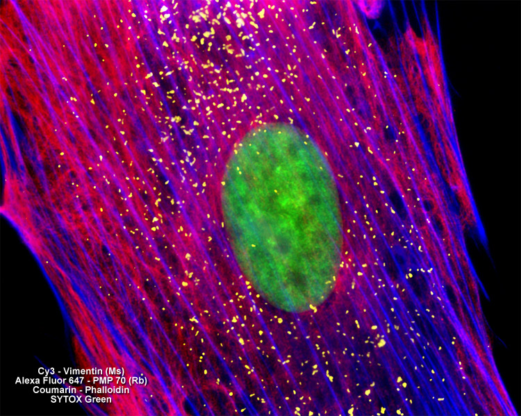
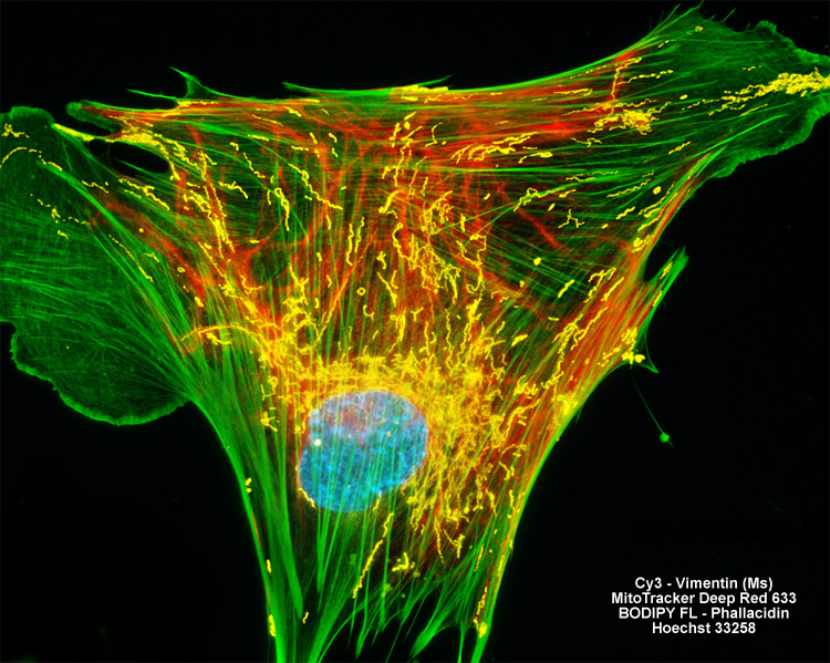

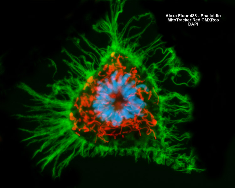
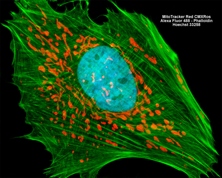
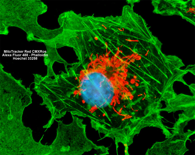
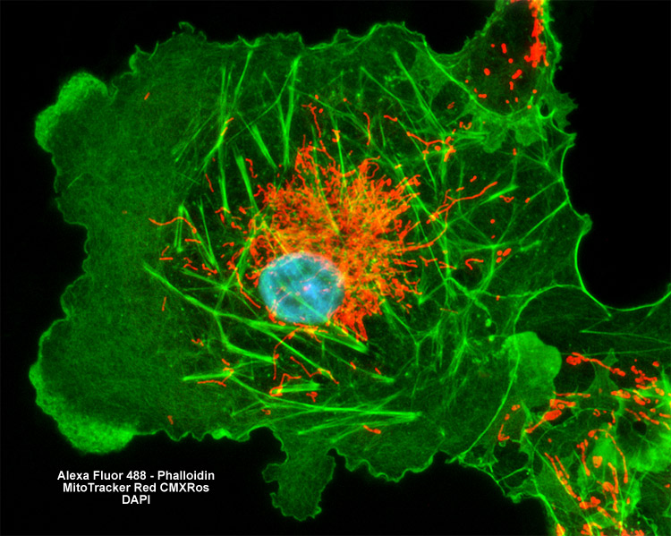
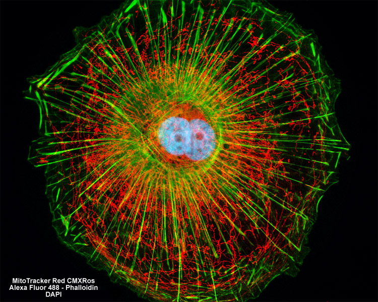
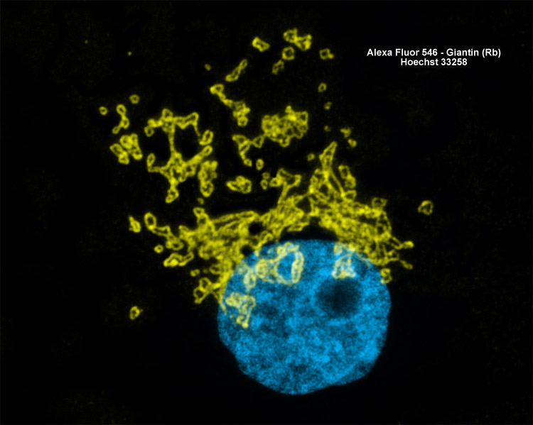
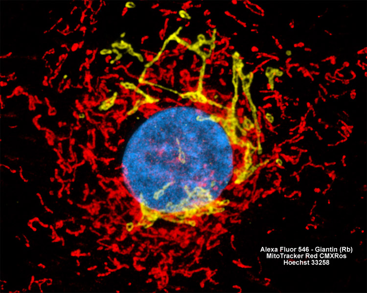
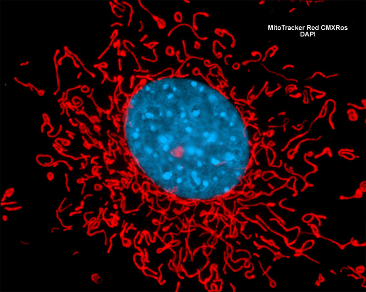

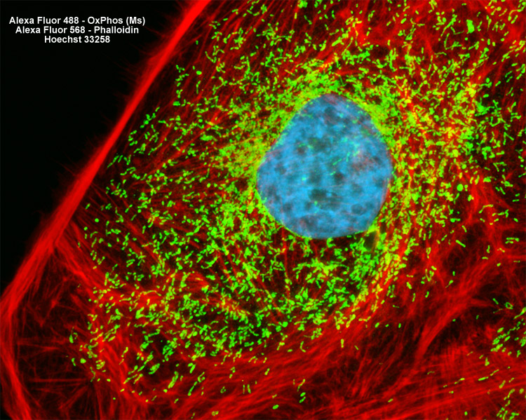
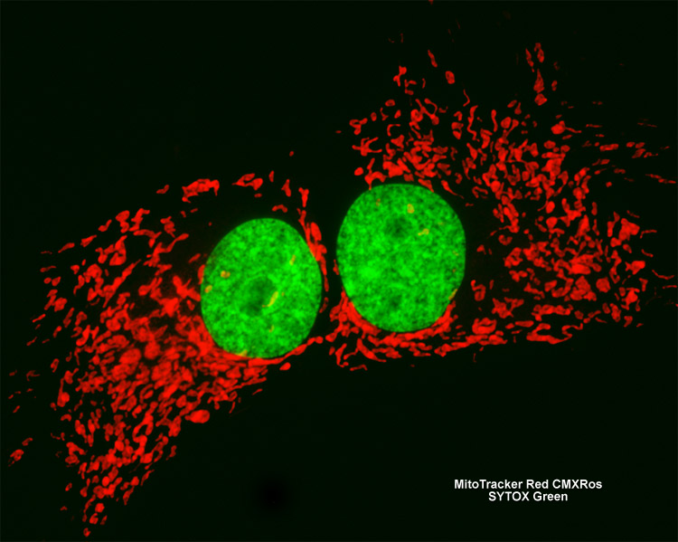
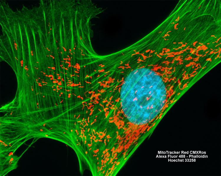
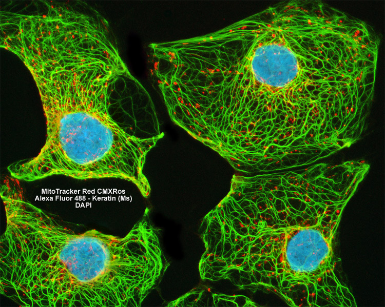
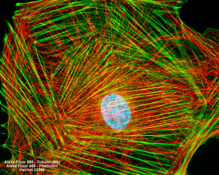
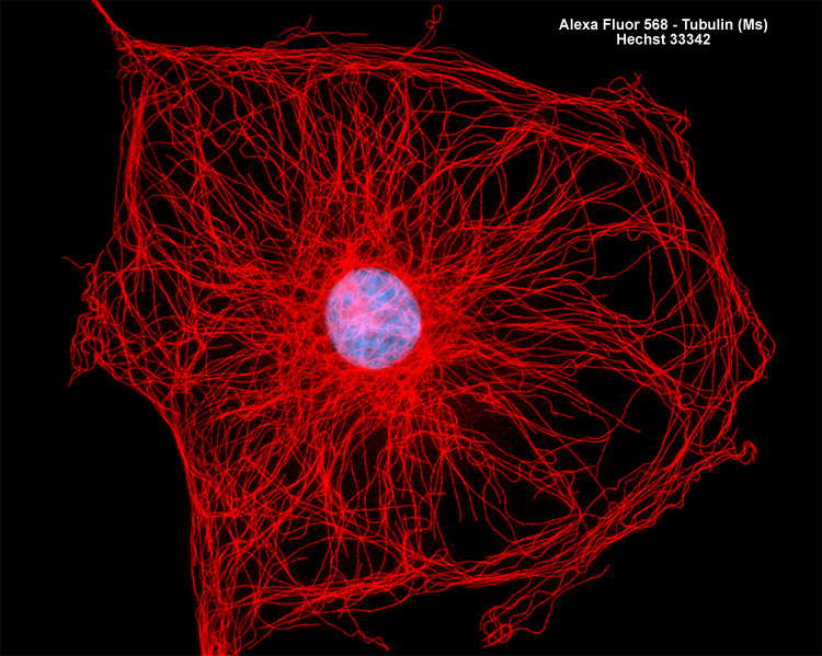
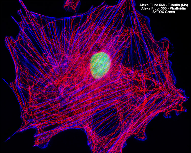
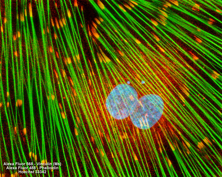

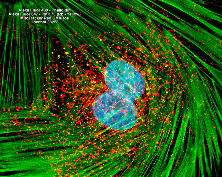

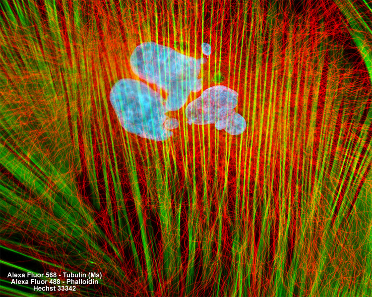

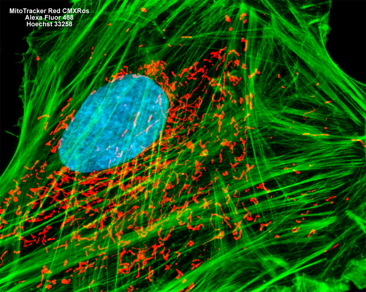
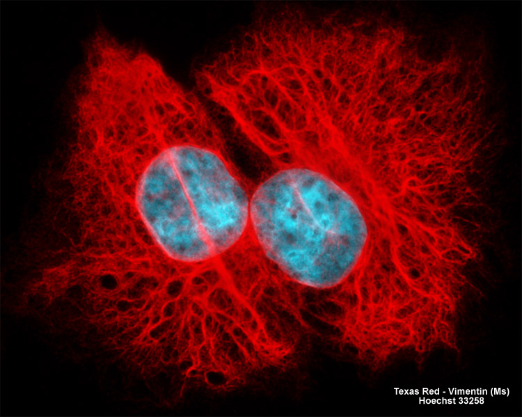
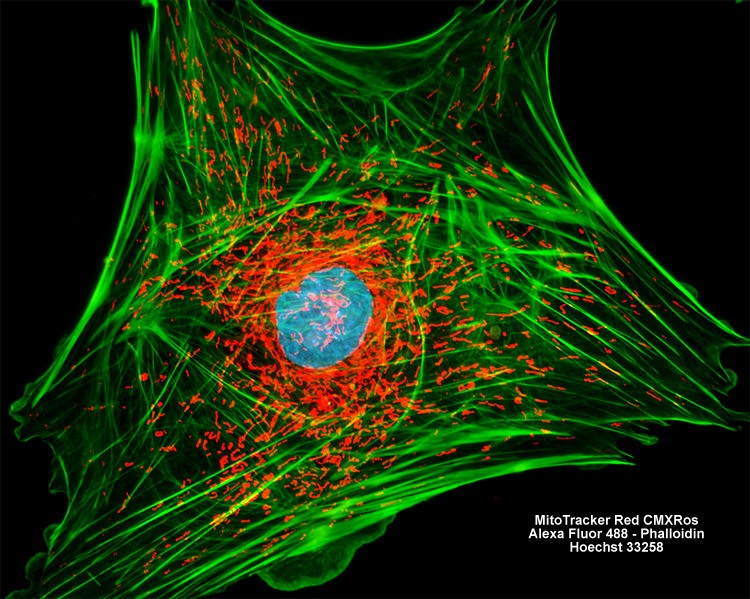
The digital images in this gallery explore multiple staining using a variety of cell lines labeled with fluorophores that target specific organelles. Images were produced using a ZEISS AxioImager upright microscope equipped with filter sets for ultraviolet, cyan, green, and yellow excitation with bandpass emission.