


Mixed Pollen - 63x (Autofluorescence)
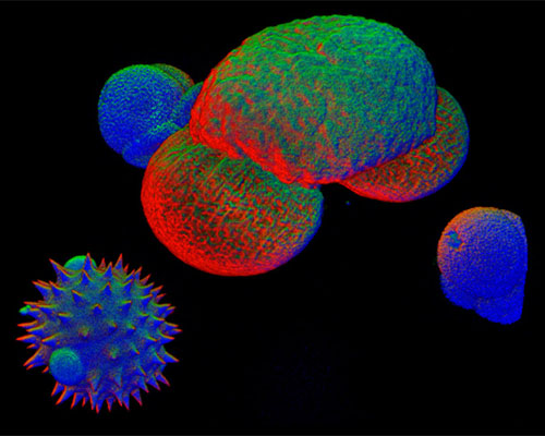
The digital image above is a reconstruction of thick optical sections of a sample of mixed pollen imaged by means of autofluorescence. To the naked eye, pollen typically appears as a fine dust, but the dust is composed of thousands of tiny grains. Pollen grains vary greatly in size, shape, and surface patterns. Because of the tremendous variety that occurs, scientists can often use these characteristics as indicators of plant genus or species. The basic structure of pollen, which includes both an inner (intine) and outer (exine) wall surrounding the male gamete, makes the grains exceptionally resilient and resistant to decay.