


Mouse Bladder Tissue - 40x
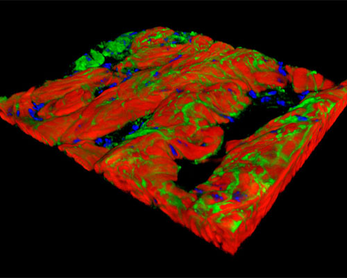
Shown above is a three-dimensional reconstruction of mouse bladder tissue stained with Alexa Fluor 350 (wheat germ agglutinin; lectins), Alexa Fluor 568 (phalloidin; actin), and SYTOX Green (nuclei). The bladder is a hollow, distensible organ that primarily serves to collect the urine excreted by the kidneys. The bladder wall is comprised of multiple layers, one of which consists of the powerful detrusor muscle. In conjunction with gravity and intrabdominal pressure, the detrusor muscle triggers the emptying of the bladder. In order to characterize the detrusor muscle and better understand the spontaneous contractile behavior of the organ, scientists have often utilized the mouse bladder for research purposes.