


Mouse Brain Tissue - 20µm - 40x
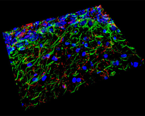
Shown above is a three-dimensional reconstruction of 20 micrometer horizontal sections of mouse brain tissue that were fixed and stained with Alexa Fluor 405 (histones), Alexa Fluor 488 (neurofilaments), and Alexa Fluor 568 (GFAP). The mouse brain has proven to be extremely useful as a model for revealing details of the complex anatomy and physiology of the human brain. Through studies of the mouse brain, researchers have also learned a great deal about serious maladies that affect the brain, such as Parkinsons, Alzheimers, and Lou Gehrigs disease. As a better understanding of such diseases is obtained, there is increasing hope for enhanced treatments and possible cures for the wide variety of brain disorders that account for one-third of all chronic illnesses.