


 African Green Monkey Kidney Fibroblast Cells")
A derivative line initiated from the CV-1 African green monkey kidney cell line, COS-1 was produced by transformation of the earlier line with an origin defective mutant of simian virus 40 (SV40) that codes for wild type T-antigen. COS-1 cells contain a single integrated copy of the complete early region of SV40 DNA.
Transformed (Simian Virus 40) African Green Monkey Kidney Fibroblast Cells
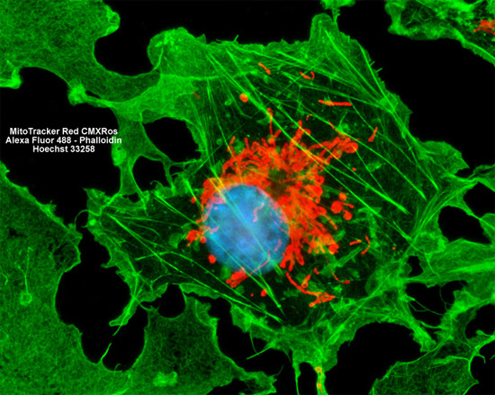
The COS-1 monkey kidney cells featured in the digital image above were resident in a culture fluorescently labeled for mitochondria with MitoTracker Red CMXRos and for the filamentous actin network with Alexa Fluor 488 conjugated to phalloidin. Cell nuclei were visualized with a blue nucleic acid stain (Hoechst 33258). Images were recorded in grayscale with a Hamamatsu ORCA AG camera system coupled to a ZEISS Axio Imager microscope equipped with bandpass emission fluorescence filter optical blocks provided by Chroma and Semrock. During the processing stage, individual image channels were pseudocolored with RGB values corresponding to each of the fluorophore emission spectral profiles.