


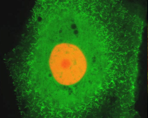
Pig Kidney Cells with mEmerald-Fascin and mCherry-H2B
The LLC-PK1 cell line is derived from the normal male pig kidney (epithelial morphology) that was 3 to 4 weeks of age. Cellular products include tissue plasminogen activator (tPA). The cell line is used for pharmacologic and metabolic studies as well as molecular cloning.
The pig kidney epithelial cells (LLC-PK1 line) featured in the digital video sequences in this section are shown expressing mEmerald fused to fascin and mCherry fused to H2B.
The green fluorescent protein fused to fascin, mEmerald, is a derivative of EGFP selected for improved brightness and photostability. Peak excitation and emission of mEmerald occur at 487 and 509 nanometers, respectively. The red fluorescent protein employed to visualize H2B, mCherry, is an original member of the monomeric mFruit series of fluorescent proteins developed via the directed evolution of mRFP1. Excitation and emission maxima of mCherry occur at 587 and 610 nanometers, respectively.