


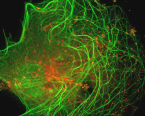
Pig Kidney Cells with mEmerald-MAP4 and mCherry-Rab5a
mCherry, in comparison with other monomeric red photoactivatable proteins, has better pH stability, faster maturation and photoactivation, higher photoactivation contrast and improved photostability. Its green fluorescence deficiency and single-molecule behavior make monomeric PAmCherry1 a favored tag for super-resolution techniques and for two-color diffraction-limited photoactivation imaging.
The physical relationship between early endosomes and the microtubule network is explored in this digital video sequence. Endosomes in cultured pig kidney epithelial cells (LLC-PK1 line) were visualized with mCherry fused to Rab5a and microtubules were fluorescently labeled with mEmerald fused to MAP4. mCherry is a red member of the mFruit series of fluorescent proteins developed via mutagenesis of mRFP1. Excitation and emission of mCherry peak at 587 and 610 nanometers, respectively. mEmerald is a green, high-performance variant of the enhanced Aequorea derivative EGFP. Peak excitation of mEmerald occurs at 487 nanometers and emission of the fluorescent protein peaks at 509 nanometers.