


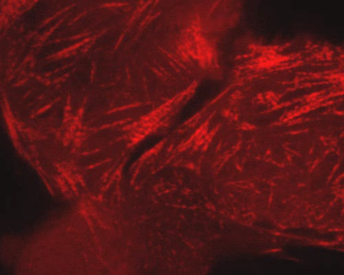
Human Osteosarcoma with mRuby-LC Myosin
Mysoins are an important group of molecular motor proteins present in nearly all eukaryotic cells. The main characteristics held in common by the various known myosin isoforms include actin-binding, force transducing, and ATP hydrolyzing capabilities. Most myosins are comprised of three domains: head, neck, and tail. The neck domain is the region that often serves as a binding site for light chain myosins, which are distinct proteins that form part of a macromolecular complex with other light chain and heavy chain myosins. Light chain myosins often function in a regulatory capacity.
Cultured human osteosarcoma cells (U-2 line) appear in this digital video sequence expressing a red fluorescent protein tag fused to light chain myosin. mRuby is a derivative of eqFP611, a far-red fluorescent protein derived from the bubble-tip anemone (Entacmaea quadricolor). Excitation and emission of mRuby peak at 558 and 605 nanometers, respectively.