


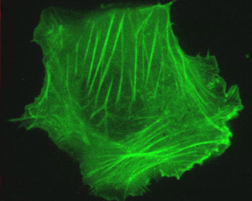
Human Osteosarcoma with mTag-GFP2 and Actin
Actin is an abundant cellular protein that plays a fundamental role in cell structure and motility. The protein exists both as a monomer (G-actin) and in lengthy, polymerized rods (F-actin) termed microfilaments. A number of actin-binding proteins have been identified that influence actin polymerization and depolymerization, as well as the bundling of microfilaments. Profilin, for example, is a eukaryotic protein that can bind to G-actin to preclude the polymerization of actin.
Actin was visualized in the human osteosarcoma cells (U-2 line) featured in this digital video sequence with mTagGFP2. A monomeric green fluorescent protein, mTagGFP2 is a variant of a mutant of the Aequorea macrodactyla GFP-like fluorescent protein. mTagGFP2 is roughly equivalent to GFP in terms of brightness, but mTagGFP2 exhibits significantly improved photostability. Excitation and emission of mTagGFP2 peak at 483 and 506 nanometers, respectively.