


EGFP Fused to Human beta-Catenin
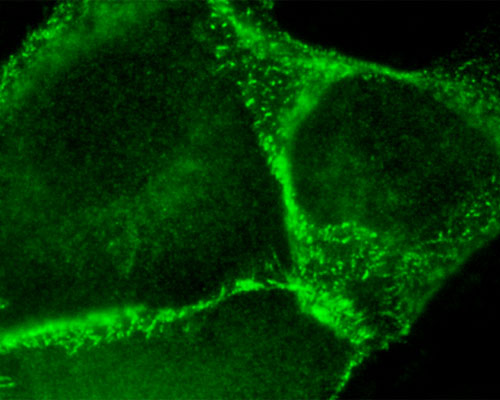
The digital image above shows mammalian cells expressing the Aequorea victoria-based protein, EGFP, fused to beta-catenin. This fluorescent protein, which possesses an excitation spectral profile well suited to the 488-nanometer argon ion laser line, can be seen localized in areas where neighboring cells come into contact with one another. This is because members of the catenin family of peripheral cytosolic proteins bind selectively to the highly conserved cytoplasmic tail domain of the cell-to-cell adhesion cadherin proteins. beta-catenin, which is one of three major catenin species, binds directly to the E-cadherin cytoplasmic domain and is crucial to intercellular adhesion and signal transduction between adjacent cells.