


mKate2 Fused to LAMP1
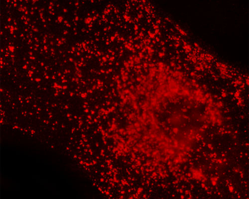
mKate is a monomeric far-red fluorescent protein with excellent photostability that is only about one-fourth as bright as EGFP. The peak excitation and emission wavelengths of mKate are 588 nanometers and 633 nanometers, respectively. Through continued engineering of mKate, a significantly brighter variant with similar spectral characteristics called mKate2 was developed. In the digital image above, the localization of mKate2 fused with lysosomal-associated membrane protein 1 (LAMP1) is demonstrated. As can be observed, lysosomes are roughly spherical microbodies located throughout the cell interior. These organelles primarily function in the digestion of various cellular materials, such as waste products, proteins, fats, and carbohydrates.