


mVenus Fused to Mouse Talin 1
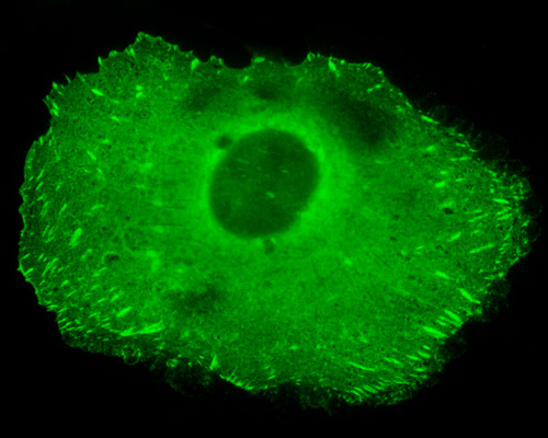
Native Aequorea victoria GFP and its derivatives, such as its enhanced form known as EGFP, exhibit a weak tendency to dimerize. Unfortunately, the formation of dimers and higher order oligomers can be problematic for cell biologists because they can produce atypical localization. In order to avoid this problem, researchers developed a method to convert the fluorescent proteins into monomers using one of several point mutations with A206K (lysine substituted for alanine at amino acid position 206). The monomeric variant of Venus (a yellow fluorescent protein), mVenus, can be seen fused to mouse talin 1 in the cell sample appearing in the above digital image. Talins are cytoskeletal proteins that localize in regions of cell-substratum contact, or in the case of lymphocytes, in sites of cell-cell contact. Studies indicate that talins interact with a variety of proteins, such as integrins, vinculin, and alpha-actinin.