


mCherry Fused to Human gamma-Tubulin
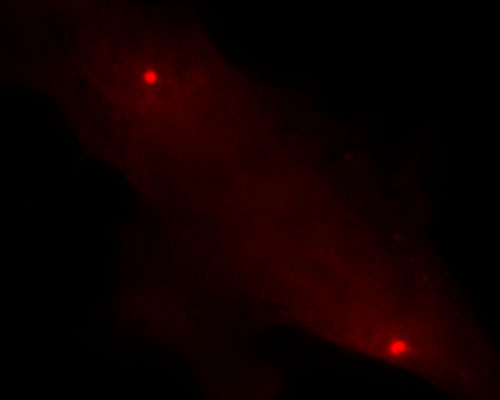
The two primary components of microtubules are alpha-tubulin and beta-tubulin, but there are several other less common members of the tubulin protein family as well. One of them, termed gamma-tubulin, resides chiefly in centrosomes and spindle pole bodies. gamma-tubulin plays a key role in microtubule organization, forming ring-shaped complexes with proteins that are thought to act as templates for the nucleation of microtubules. The localization of a fusion of mCherry to gamma-tubulin is demonstrated in the digital image above. Like the other "mFruits", mCherry was developed from the first generation red fluorescent protein (mRFP1) through a directed evolution approach. The peak excitation and emission wavelengths of mCherry are 587 and 610 nanometers, respectively.