


Mouse Brain Tissue
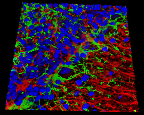
Presented above is a three-dimensional reconstruction of a 16-micrometer section of mouse cerebellum tissue that was fixed with paraformaldehyde and stained with Alexa Fluor 488 (neurofilaments), Alexa Fluor 405 (histones), and Alexa Fluor 568 (GFAP). The mouse brain has proven to be exceptionally useful as a model for revealing details of the intricate anatomy and physiology of the human brain. Through studies of the mouse brain, researchers have discovered many details about serious maladies that affect the brain, such as Parkinson's, Alzheimer's, and Lou Gehrig's disease.