


Mouse Brain Tissue
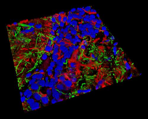
Featured in the above digital image is a three-dimensional horizontal reconstruction of a 20-micrometer section of mouse brain tissue that was stained with Alexa Fluor 488 (neurofilaments), Alexa Fluor 568 (phalloidin; actin), and GFAP (glial fibrillar acidic protein). High resolution imaging of mouse brain activity is becoming increasingly important in the pursuit of insight into human neurological disease. As more information from these diseases is acquired, there is greater hope for enhanced treatments and possible cures for the wide range of brain disorders that are accountable for one-third of all chronic illnesses.