


Mouse Colon Tissue
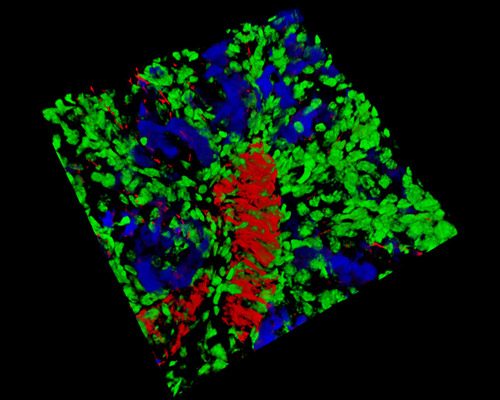
This digital image in this section is a three-dimensional reconstruction of a mouse colon tissue sample that was stained with Alexa Fluor 350 (wheat germ agglutinin; highlighting lectins), Alexa Fluor 568 (phalloidin; labeling actin filaments), and SYTOX Green (nuclei). With 639,000 deaths worldwide per year, colorectal cancer is the third leading cause of cancer-related death. The abnormal tube-like glands that can develop into cancerous lesions are called Aberrant Crypt Foci (ACF). As in humans, the crypts (that renew the lining of the intestine and make mucus) that undergo morphological changes are known in mice to be consistent biomarkers for colon cancer and are commonly used to study initiation, promotion, and chemoprevention of colorectal cancer.