


Mouse Colon Tissue
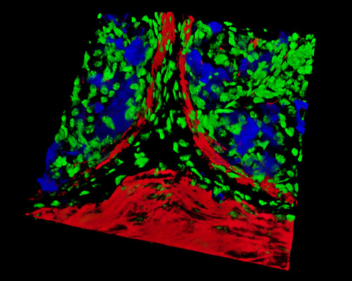
In the digital image featured in this section is a three-dimensional reconstruction of a mouse colon tissue sample that was stained with Alexa Fluor 350 (wheat germ agglutinin; highlighting lectins), Alexa Fluor 568 (phalloidin; labeling actin filaments), and SYTOX Green (nuclei). The colon, cecum, and rectum structure the large intestine. In mice and in other mammals, the colon is comprised of four regions: the ascending, transverse, descending, and sigmoid colon.