


Mouse Stomach Tissue
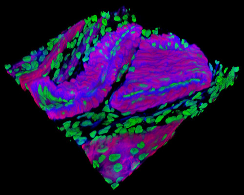
Featured in this section is a three-dimensional reconstruction of mouse stomach tissue stained with Alexa Fluor 350 (wheat germ agglutinin; highlighting lectins), Alexa Fluor 568 (phalloidin; labeling actin filaments), and SYTOX Green (nuclei). The layer of epithelial cells directly exposed to the stomach's acidic, gastric lumen is the most jeopardized component of the gastric mucosa, as gastric pit lumens have been found to have the most divergent pH from luminal superfusates. Mice and other mammals as well as amphibian models are preferential for examining gastric repair procedures because they can provide physiological significant response for understanding similar events in human ulcer disease.