


Rat Brain Tissue
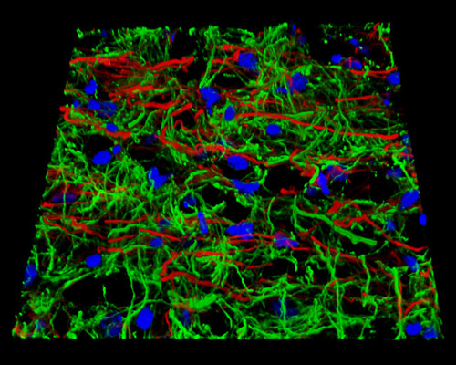
The digital image featured above is a three-dimensional reconstruction of a 16-micrometer horizontal section of rat brain tissue stained with Alexa Fluor 488 (WGA), Alexa Fluor 568 (phalloidin; labeling actin filaments), and Alexa Fluor 350 (WGA). Much of what we know about the human brain has been revealed from research on the rat brain. The rat brain is exceptionally useful as a subject of study, though smaller and less complex than that of humans, because most regions of the brain are fundamentally similar among mammals. The rat brain has been studied as a model for a range of neurological diseases, such as Parkinson's disease. In fact, through studying rats, the breaking discovery that Parkinson's is caused by the loss of dopamine within the brain was made. Research with the rodents has also been vital in testing new treatments for the disease, as well as examinations of other potential therapeutic approaches, such as gene therapy.