


Rat Diaphragm Tissue
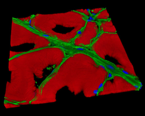
In the digital image presented in the above section, a 16-micrometer section of rat diaphragm tissue is featured in a 3D reconstruction. The sample is stained with Alexa Fluor 488 (neurofilaments), Alexa Fluor 568 (phalloidin; labeling actin filaments), and DAPI (nuclei). The muscular fibers of the diaphragm are divided into three parts: the sternal, which is the small slip on each side that comes from the inner surface of the xiphoid process and inserts on the central tendon; the costal, which comes up from the inner aspect of the lower six costal cartilages and the lower four ribs and plants on to the anterolateral part of the central tendon; and the lumbar, the portion that rises from the upper lumbar vertebrae and from the medial and lateral arcuate ligaments.