


Rat Esophagus Tissue
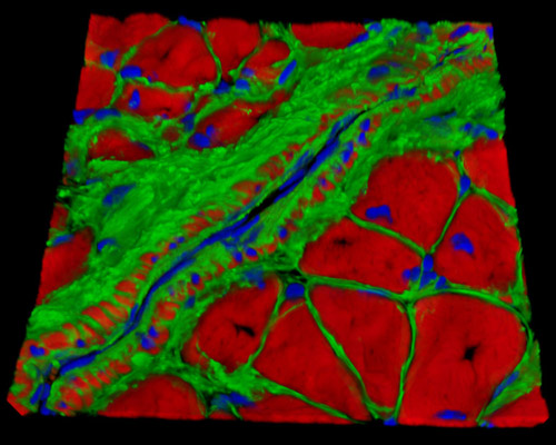
The digital image presented above is a three-dimensional reconstruction of a 16-micrometer section of rat esophagus tissue stained with Alexa Fluor 488 (wheat germ agglutinin; highlighting lectins), Alexa Fluor 568 (phalloidin; labeling actin filaments), and DAPI (nuclei). Andocarcinoma of the esophagus is the most common malignant tumor of the esophagus. In rats with esophagogastroduodenal anastomosis (EGDA), going without treatment can lead to columnar-lined esophagus (CLE) including esophageal adenocarcinoma (EAC). EGDA rats may prove as a useful model to study the pathogenesis, molecular biology, and chemo-preventive interventions of human Barrett's esophagus (the abnormal growth of intestinal-type cells in the inferior portion of the esophagus) and EAC.