


Rat Jejunum Tissue
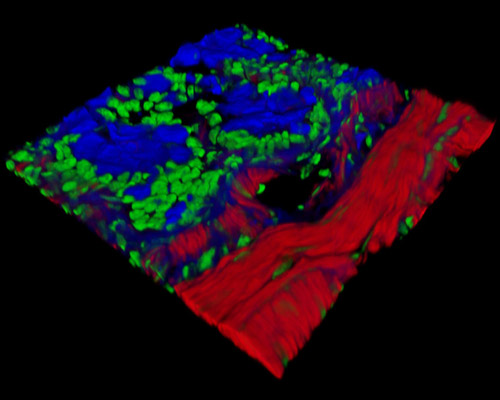
The digital image featured above is a 16-micrometer section of a three-dimensional reconstruction of rat jejunum tissue stained with Alexa Fluor 350 (wheat germ agglutinin; highlighting lectins), Alexa Fluor 568 (phalloidin; labeling actin filaments), and SYTOX Green (nuclei). The jejunum is the second of three major sections (the duodenum, jejunum, and ileum) of the small intestine present in mammals and the majority of higher vertebrates. It accepts digested material from the duodenum through peristalsis, which is facilitated by inner circular and outer longitudinal layers of smooth muscle along the duodenum and much of the gastrointestinal tract.