


Rat Jugular Tissue
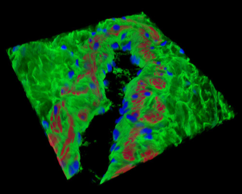
The three-dimensional reconstruction featured above depicts a 16-micrometer section of a rat jugular vein stained with Alexa Fluor 488 (wheat germ agglutinin; highlighting lectins), Alexa Fluor 568 (phalloidin; labeling actin filaments), and DAPI (nuclei). The jugular veins bring deoxygenated blood from the head to the heart through the superior vena cava. The internal jugular is contained inside the carotid sheath with the common carotid artery and vagus nerve, providing venous drainage for the skull's contents. The external jugular receives most of the blood from the exterior of the cranium and the deep sections of the face.