


Rat Stomach Tissue
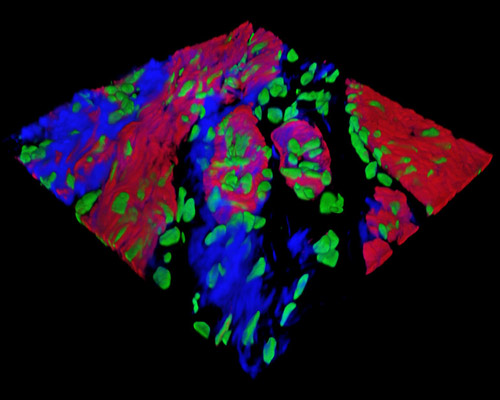
Presented in the digital image above is a three-dimensional reconstruction of a 16-micrometer section of rat stomach cardiac tissue that was stained with Alexa Fluor 488 (wheat germ agglutinin; lectins), Alexa Fluor 568 (phalloidin; actin), and SYTOX Green (nuclei). The distal wall of the groove between the rat forestomach and glandular stomach is lined with fibrillovesicular cells (FVC) that are characterized by microvilli, microfilaments, and a complex tubulovesicular system. The wall is also lined with a specific type of columnar cells (CCGG) that contain small mucous granules and special vesicles and tubules. The cardiac glands contain cardiac mucous (CMC), which are filled with large mucous granules and resemble mucous neck cells and serous cells (CSC).