


Rat Vena Cava Tissue
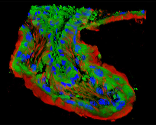
The digital image in this section depicts a three-dimensional reconstruction of rat vena cava tissue stained with Alexa Fluor 350 (WGA; highlighting lectins), Alexa Fluor 568 (phalloidin; labeling actin filaments), and SYTOX Green (nuclei). Entering the right atrium are three main blood vessels that bring the deoxygenated blood back to the heart from all regions of the body. These blood vessels are the right superior vena cava, the left superior vena cava, and the inferior vena cava. The right and left superior venae cavae return deoxygenated blood to the heart from the right and left side of the head and neck. The inferior returns deoxygenated blood to the heart from the lower part of the body.