


Rat Vena Cava Tissue
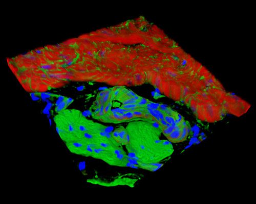
Presented in this section is a three-dimensional reconstruction of rat vena cava tissue stained with Alexa Fluor 488 (WGA; highlighting lectins), Alexa Fluor 568 (phalloidin; labeling actin filaments), and DAPI (nuclei). Superior vena cava syndrome (SVCS) is characterized by gradual, insidious compression of the superior vena cava. Because the syndrome is often typified by a slow increase in symptomatology, diagnosis is often delayed until major compression of the superior vena cava has occurred. Rats have two superior venae cavae, however, in contrast to humans, who only have one. Rats' possession of the left superior vena cava allows the cerebral veins to decompress and minimize the occurrence of cerebral edema, the excess accumulation of water in the intracellular and/or extracellular spaces of the brain, which is sometimes postoperatively caused by the acute rise in superior vena cava pressure.