


Mouse Embryo Tissue Section
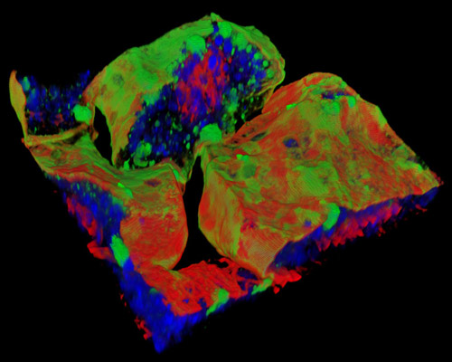
The digital image depicted in this section is a three-dimensional reconstruction of a 30-micrometer section of mouse embryo tissue at 17 days stained with Alexa Fluor 488 (wheat germ agglutinin; highlighting lectins), Alexa Fluor 568 (phalloidin; labeling actin filaments), and DAPI (nuclei). For more than half a decade, it has been recognized that folate, a water-soluble B vitamin that must be attained through diet or supplementation, plays an essential role in embryonic development. Research has proved that women who had a fetus with a neural tube defect also had blood levels and diets that were deprived of many micronutrients, but particularly folate. Inactivation of genes in the folate pathway of mice results in malformations of the heart, neural tube, and craniofacial structures, which conclusively led to the United States Public Health Service recommendation that all pregnant women, not only those with previously affected offspring, take folic acid supplements.