


Mouse Embryo Tissue Section
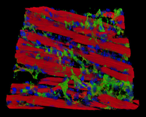
In the digital image presented in this section, a 30-micrometer section of mouse embryo tissue at 17 days is presented in a 3D reconstruction. The sample is stained with Alexa Fluor 488 (wheat germ agglutinin; highlighting lectins), Alexa Fluor 568 (phalloidin; labeling actin filaments), and DAPI (nuclei). An embryo can be assigned a Carnegie stage (a number 1 to 23) by embryologists to describe its perceptible maturity based on its external features, such as eyes, nose, ribs, or mouth. Postovulatory age is used by clinicians to describe the maturity of an embryo based on the amount of time since the last ovulation before pregnancy.