


Mouse Embryo Tissue Section
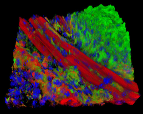
The digital image depicted in this section is a three-dimensional reconstruction of a 30-micrometer section of mouse embryo tissue at 17 days stained with Alexa Fluor 488 (wheat germ agglutinin; highlighting lectins), Alexa Fluor 568 (phalloidin; labeling actin filaments), and DAPI (nuclei). Mice are one of few species of mammals that have produced self-renewing embryonic stem cells. The methods for growing mouse embryonic stem cells from the inner cell accumulation of the preimplantation blastocyst were first reported almost 30 years ago, and now, versions of the procedure are used in laboratories throughout the world. Studies of embryonal carcinoma cells from mice have also helped launch parameters for growing and evaluating embryonic stem cells.