


Mouse Embryo Tissue Section
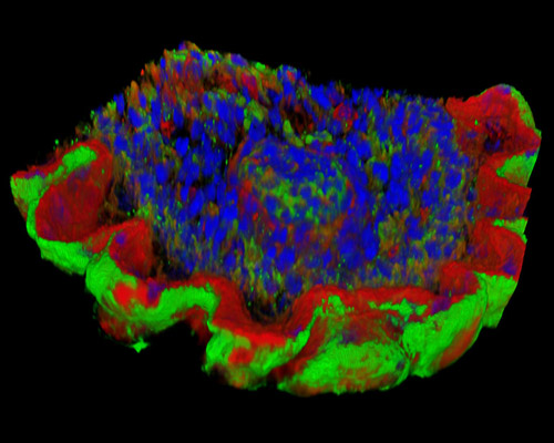
The digital image depicted in this section is a three-dimensional reconstruction of a 30-micrometer section of mouse embryo tissue at 17 days stained with Alexa Fluor 488 (wheat germ agglutinin; highlighting lectins), Alexa Fluor 568 (phalloidin; labeling actin filaments), and DAPI (nuclei). Until an early post-implantation stage at least, pluriopotential cells are present in mouse embryo, as revealed by their part in the formation of chimeric animals and ability to form teratocarcinomas. It has recently become possible to produce growing cultures of these cells in vitro, and cell lines can now be directly isolated from in vitro cultures of mouse blastocysts, leading to further advances in cancer research.