


Mouse Embryo Tissue Section
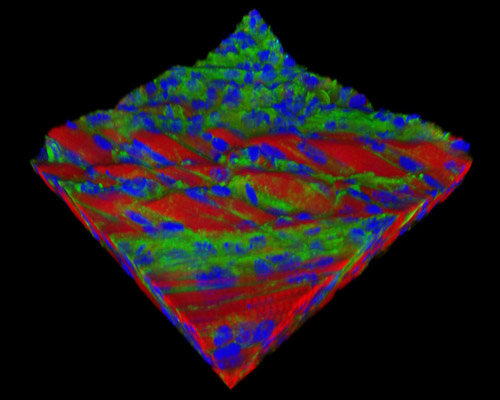
The digital image featured in this section depicts a three-dimensional reconstruction of a 30-micrometer section of mouse embryo tissue at 17 days stained with Alexa Fluor 488 (wheat germ agglutinin; highlighting lectins), Alexa Fluor 568 (phalloidin; labeling actin filaments), and DAPI (nuclei). In order to reduce the developmental variation of mice embryos, the Theiler and Carnegie staging systems can be used. Up to Theiler stage 19, each phase covers one half day of gestation, but the succeeding stages cover about one day of gestation each.