


Mouse Embryo Tissue Section
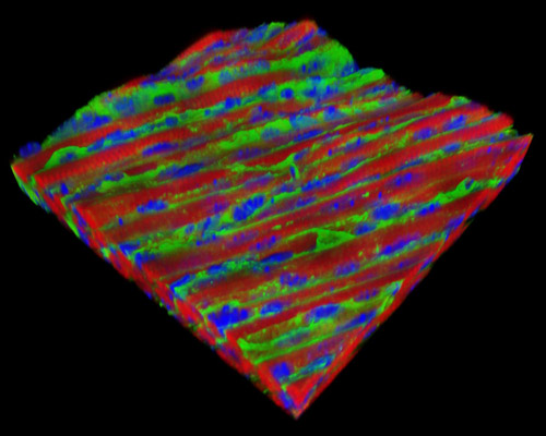
Featured in this section is a three-dimensional reconstruction of mouse embryo tissue at 17 days stained with Alexa Fluor 488 (wheat germ agglutinin; highlighting lectins), Alexa Fluor 568 (phalloidin; labeling actin filaments), and DAPI (nuclei). In the last several stages of the Theiler system, the rodent embryo is undergoing its final developments. Fingers and toes separate, become parallel, and the eyelids close. The skin thickens and forms wrinkles, the subcutaneous veins are less visible, and the umbilical hernia disappears. Long whiskers become apparent, and the eyes become visible through closed eyelids before finally, a new born rodent goes through post-natal development.