


Rat Embryo Tissue Section
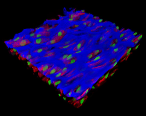
The digital image presented in this section is a three-dimensional reconstruction of a 30-micrometer section of rat embryo tissue at 19 days that was stained with Alexa Fluor 350 (wheat germ agglutinin; highlighting lectins), Alexa Fluor 568 (phalloidin; labeling actin filaments), and SYTOX Green (nuclei). Some of the advantages of using the rat embryo model in contrast to the mouse embryo model include the rat model being much larger, easier to breed, and in vision studies, the rat is preferable because the retinal development continues postnatally, whereas most other vertebrate neurological systems are hard to access during periods of development. Rat development is also typically 2 days behind that of a mouse.