


Rat Embryo Tissue Section
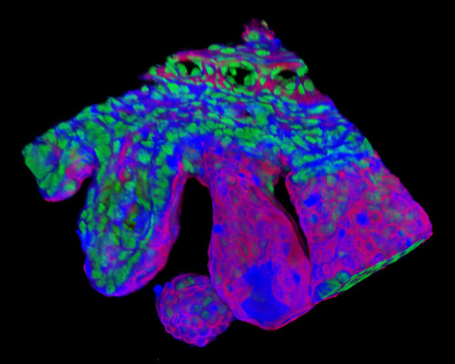
The digital image featured in this section depicts a three-dimensional reconstruction of a 30-micrometer section of rat embryo tissue at 19 days stained with Alexa Fluor 350 (wheat germ agglutinin; highlighting lectins), Alexa Fluor 568 (phalloidin; labeling actin filaments), and SYTOX Green (nuclei). On day 19 of a rat's development, the embryo is near the end of its transformation. It is at least in its first fetal stage, which is characterized by rapid growth of the eyelids, completion of the palate, the withdrawal of the umbilical hernia, and the pinna covering the ear duct.