


Rat Embryo Tissue Section
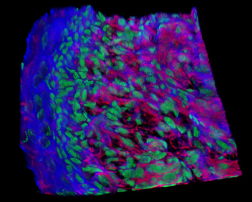
Featured in this section is a three-dimensional reconstruction of rat embryo tissue at 19 days stained with Alexa Fluor 350 (wheat germ agglutinin; highlighting lectins), Alexa Fluor 568 (phalloidin; labeling actin filaments), and SYTOX Green (nuclei). Much of what we know about human embryonic development is derived from studies of embryogenesis in the rodent. Several facets of embryogenesis in mice are similar to that of humans, but development in mice is also different from human development in several ways. For instance, embryonic and fetal development in mice takes 18 to 20 days, whereas in humans, the course of development takes nine months.