


Rat Embryo Tissue Section
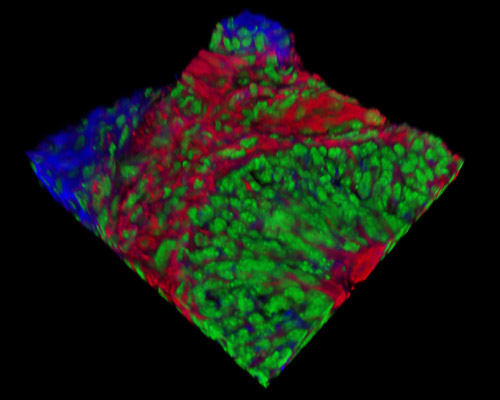
Presented in the digital image in this section is a three-dimensional reconstruction of rat embryo tissue at 19 days stained with Alexa Fluor 350 (wheat germ agglutinin; highlighting lectins), Alexa Fluor 568 (phalloidin; labeling actin filaments), and SYTOX Green (nuclei). The placenta forms and functions differently in mice and in humans. In mice, an egg cylinder forms after the embryo implants in the uterine wall; in humans, an embryonic disk develops. The yolk sac of a rodent embryo lasts and functions throughout gestation, whereas in humans, it functions in early embryogenesis alone.