


Rat Embryo Tissue Section
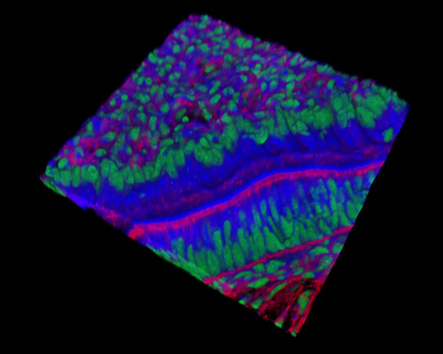
The digital image presented in this section is a three-dimensional reconstruction of a 30-micrometer section of rat embryo tissue at 19 days that was stained with Alexa Fluor 350 (wheat germ agglutinin; highlighting lectins), Alexa Fluor 568 (phalloidin; labeling actin filaments), and SYTOX Green (nuclei). As the embryo forms, its overall body pattern is determined by the growth of three specific body axes: the anterior-posterior axis (head-tail), the dorsal-ventral (back-belly) axis, and left-right asymmetry. These axes must be established at the correct time, as timing is crucial to normal embryonic development.