


Rat Embryo Tissue Section
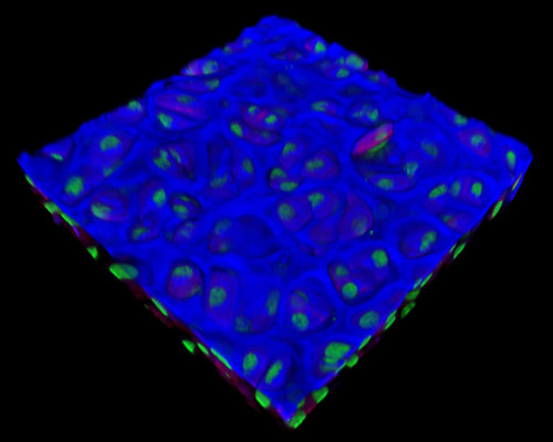
The digital image featured in this section depicts a three-dimensional reconstruction of a 30-micrometer section of rat embryo tissue at 19 days stained with Alexa Fluor 350 (wheat germ agglutinin; highlighting lectins), Alexa Fluor 568 (phalloidin; labeling actin filaments), and SYTOX Green (nuclei). Several cells of the embryo in its beginning stages are in a continuous state of dividing or of preparing to divide. The cell cycle is the progression of molecular events that regulate these processes. It includes four major phases: DNA synthesis (S phase), G2 (a gap phase in which the cell size increases and prepares to divide), cell division (mitosis), and G1 (replication of the centrioles and a gap phase of cell growth).