


Rat Embryo Tissue Section
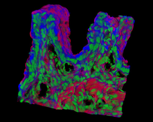
Presented in the digital image in this section is a three-dimensional reconstruction of rat embryo tissue at 19 days stained with Alexa Fluor 350 (wheat germ agglutinin; highlighting lectins), Alexa Fluor 568 (phalloidin; labeling actin filaments), and SYTOX Green (nuclei). Embryonic stem cells have been available from inbred mice since 1981, but it was not until 2008 that rat ES cells were reportedly derived, important in that the rat's larger size makes it the model species of choice for several features of biomedical research. Toxicology studies, physiological interventions, and assessment of higher-order functions such as cognition and behavior are generally more refined and revealing in rats than in mice. The availability of rat ES cells widens the capacity for testing the ability of in vitro ES cell differentiation to create cells that can assimilate and function in adult tissue regeneration and repair.