


Rat Embryo Tissue Section
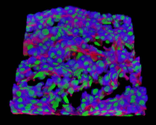
In the digital image presented in this section, a 30-micrometer section of rat embryo tissue at 19 days is presented in a 3D reconstruction. The sample is stained with Alexa Fluor 350 (wheat germ agglutinin; highlighting lectins), Alexa Fluor 568 (phalloidin; labeling actin filaments), and SYTOX Green (nuclei). The embryo is a multicellular diploid eukaryote in its beginning stage of development, which is in humans, until around eight weeks after fertilization. Embryogenesis is the process in which a sperm fertilizes an egg, and then produces a zygote that is made of half of the two parents' DNA. The zygote begins to divide by mitosis to produce the embryo.