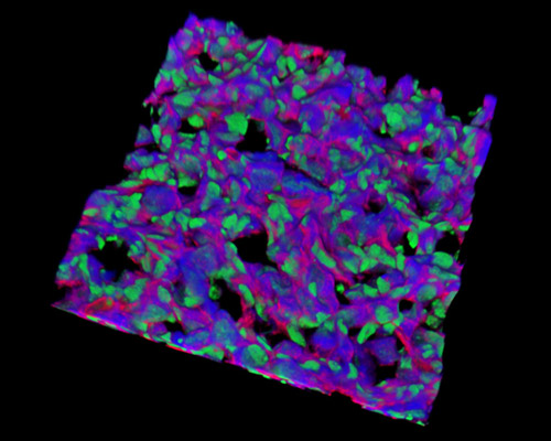
Rat Embryo Tissue Section

The 30-micrometer section presented in this digital image is a three-dimensional reconstruction of rat embryo tissue at 19 days stained with Alexa Fluor 488 (wheat germ agglutinin), Alexa Fluor 568 (phalloidin; labeling actin filaments), and DAPI (nuclei). In rats and in all animals, the growth of the zygote into an embryo ensues through three stages: the blastula stage, typified by a fluid-filled cavity called the blastocoel, which is enveloped by a mass of cells called blastomeres; the gastrula stage when the cells of the blastula experience cell division and invasion processes, and/or migrate to form a few tissue layers; and the organogenesis stage, during which the cells' developmental potential and the molecular and cellular interactions between germ layers stimulate the separation of cell types specific to the organs.


