


Rat Embryo Tissue Section
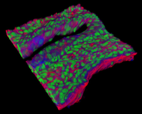
The 30-micrometer section presented in this digital image is a three-dimensional reconstruction of rat embryo tissue at 19 days stained with Alexa Fluor 350 (wheat germ agglutinin; highlighting lectins), Alexa Fluor 568 (phalloidin; labeling actin filaments), and SYTOX Green (nuclei). When embryonic development occurs in vitro or when embryonic stem cells are grown in tissue culture, normal genomic imprinting patterns can be disturbed. One way of directing mouse embryonic stem cell differentiation in vitro is to direct the stem cells to generate primitive blood vessels. Another way is to direct them to become neurons that release dopamine and serotonin. A third way is through a series of experiments that direct the differentiation of the stem cells to acquiesce pancreatic islet cells that secrete insulin.