


Rat Embryo Tissue Section
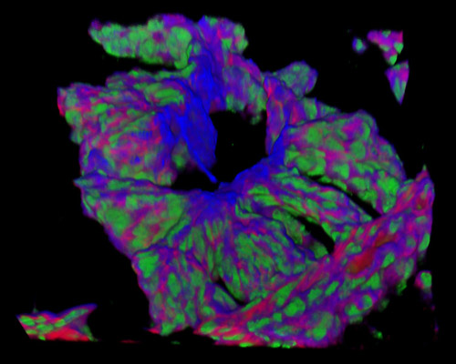
The digital image presented in this section is a three-dimensional reconstruction of a 30-micrometer section of rat embryo tissue at 19 days that was stained with Alexa Fluor 350 (wheat germ agglutinin; highlighting lectins), Alexa Fluor 568 (phalloidin; labeling actin filaments), and SYTOX Green (nuclei). The blastocyst of a lab rodent undergoes a vesicular morphology comparable to that of most other eutherian embryos, with the trophectoderm forming the vesicular wall and the inner cell mass asymmetrically located on the polar side of the blastocyst. The trophectoderm is distinguished by two entities: the polar trophectoderm related to the ICM in the embryonic compartment and the mural trophectoderm in the abembryonic cavity of the blastocyst.