


Rat Embryo Tissue Section
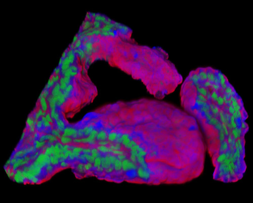
The digital image presented in this section is a three-dimensional reconstruction of a 30-micrometer section of rat embryo tissue at 19 days that was stained with Alexa Fluor 350 (wheat germ agglutinin; highlighting lectins), Alexa Fluor 568 (phalloidin; labeling actin filaments), and SYTOX Green (nuclei). The morphogenetic forces that drive the formation of the cylindrical rodent embryo are unknown. It has been hypothesized that the spatial constraint from the uterine tissues around the implanting blastocyst restricts the lateral growth of the cap of polar trophectoderm. To contain the swelling tissue mass, the polar trophectoderm must grow along the path of least resistance by jutting out into the blastocyst cavity. This results in a proximal ectoplacental cone formation and a distally extending cylindrical column of extraembryonic ectoderm.