


Rat Embryo Tissue Section
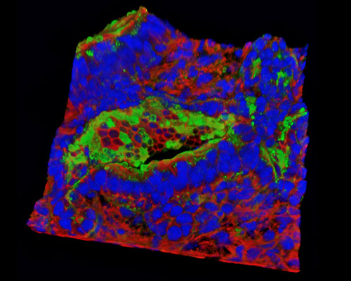
The 30-micrometer section presented in this digital image is a three-dimensional reconstruction of rat embryo tissue at 19 days stained with Alexa Fluor 488 (wheat germ agglutinin; highlighting lectins), Alexa Fluor 568 (phalloidin; labeling actin filaments), and DAPI (nuclei). Before gastrulation in rodents, the group of ICM cells is ordered into an epithelial tissue, the epiblast, which adopts the form of a cup with its rim against the distal pole of the extraembryonic ectoderm. It could be that the epiblast takes the form of a cup as a morphogenetic adaption to contain the expansion of the epithelium when the rim of the epiblast cannot enlarge sufficiently due to the limited growth in the belt of the cylindrical extraembryonic ectoderm.